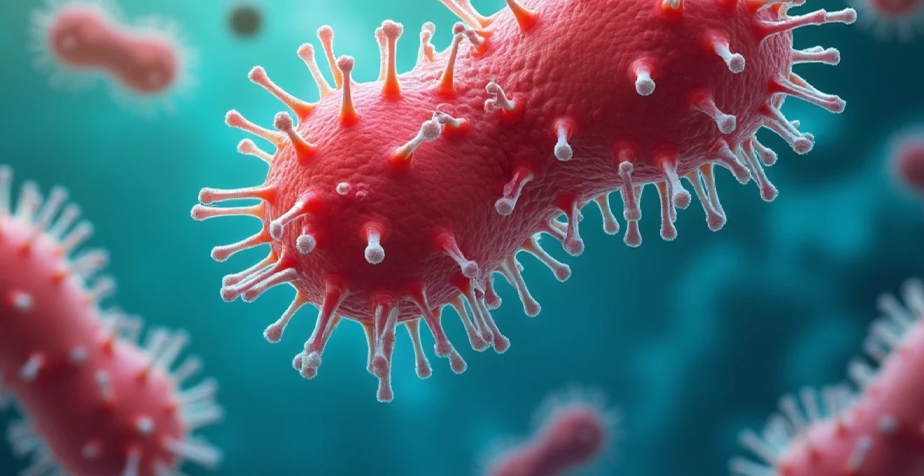
Georgia’s warm, humid climate and extensive coastal waters create ideal conditions for flesh-eating bacteria to thrive, posing significant health risks to residents and visitors alike. These aggressive pathogens, medically known as necrotising fasciitis-causing organisms, have gained attention following several high-profile cases across the state. The bacteria responsible for these devastating infections include multiple species, each with distinct characteristics and transmission patterns that healthcare professionals must understand to ensure rapid diagnosis and treatment.
The state’s unique geographical features, from the Atlantic coastline to freshwater lakes and river systems, provide diverse environments where different bacterial strains flourish. Understanding the complex interplay between environmental factors, bacterial pathogenesis, and clinical manifestations becomes crucial for both medical professionals and the public in preventing and managing these potentially fatal infections.
Necrotising fasciitis pathophysiology and bacterial classification in georgia cases
Necrotising fasciitis represents one of the most aggressive soft tissue infections, characterised by rapid tissue destruction and systemic toxicity. The pathophysiology involves bacterial invasion of deep fascial planes, where organisms produce powerful enzymes and toxins that cause widespread tissue necrosis. This process occurs at an alarming rate, often advancing several centimetres per hour, making early recognition and intervention critical for patient survival.
The infection typically begins when bacteria enter through breaks in the skin, including minor cuts, surgical incisions, or even microscopic wounds. Once established, the organisms spread along fascial planes, destroying tissue faster than the body’s immune system can respond. The bacterial toxins cause vascular thrombosis, leading to tissue ischaemia and necrosis, whilst simultaneously triggering a massive inflammatory response that can result in septic shock.
Group A streptococcus pyogenes virulence mechanisms and toxin production
Group A Streptococcus (GAS) remains the most common cause of necrotising fasciitis in Georgia, accounting for approximately 60% of cases. These bacteria possess sophisticated virulence mechanisms that enable tissue invasion and destruction. The organism produces multiple toxins, including streptolysin O and S, which damage cell membranes, and pyrogenic exotoxins that trigger toxic shock syndrome.
The M protein on the bacterial surface helps evade immune recognition whilst facilitating adherence to host tissues. Hyaluronidase, often called the “spreading factor,” breaks down connective tissue barriers, allowing rapid bacterial dissemination. Streptococcal pyrogenic exotoxin A acts as a superantigen, causing massive cytokine release that contributes to the systemic inflammatory response and shock.
Vibrio vulnificus Marine-Associated infections along georgia’s atlantic coast
Georgia’s 100-mile coastline provides optimal habitat for Vibrio vulnificus, a marine bacterium responsible for some of the most severe necrotising fasciitis cases in the state. This organism thrives in warm saltwater and brackish environments, with concentrations peaking during summer months when water temperatures exceed 20°C. The bacteria can enter the body through open wounds exposed to contaminated seawater or through consumption of raw shellfish.
Vibrio vulnificus infections progress rapidly, often requiring aggressive surgical debridement within hours of symptom onset. The organism produces cytolytic toxins that cause massive tissue destruction, whilst its polysaccharide capsule helps resist phagocytosis. Mortality rates for invasive infections can reach 50%, particularly in immunocompromised individuals or those with underlying liver disease.
Clostridium perfringens gas gangrene manifestations in freshwater environments
Clostridium perfringens, commonly found in Georgia’s freshwater systems including lakes and rivers, causes a distinct form of necrotising soft tissue infection known as gas gangrene. This anaerobic organism produces alpha toxin (phospholipase C), which destroys cell membranes and causes widespread tissue necrosis. The characteristic feature of gas gangrene includes subcutaneous emphysema, where gas bubbles become palpable under the skin.
The infection typically occurs following contamination of wounds with soil or organic matter containing spores. Lake Lanier and other recreational water bodies in Georgia have been associated with such infections, particularly following traumatic injuries sustained during water sports activities. The rapid onset and progression of symptoms, including severe pain disproportionate to physical findings, requires immediate surgical intervention.
Polymicrobial synergistic infections and mixed flora complications
Approximately 30% of necrotising fasciitis cases in Georgia involve multiple bacterial species working synergistically to cause tissue destruction. These polymicrobial infections often include combinations of aerobic and anaerobic bacteria, creating complex pathophysiological processes that complicate treatment decisions. The synergistic effect allows organisms with limited individual virulence to cause severe infections when acting together.
Common combinations include Enterobacteriaceae with anaerobic species such as Bacteroides or Peptostreptococcus. These mixed infections frequently occur in patients with diabetes, peripheral vascular disease, or following abdominal surgery. The presence of multiple organisms can lead to antibiotic resistance patterns that require broad-spectrum antimicrobial therapy and extended treatment durations.
Georgia’s environmental risk factors and endemic transmission patterns
Georgia’s environmental characteristics create a perfect storm for flesh-eating bacterial proliferation and transmission. The state’s subtropical climate, with average temperatures ranging from 16°C in winter to 27°C in summer, provides year-round conditions favourable for bacterial growth. High humidity levels, often exceeding 70%, further enhance bacterial survival in environmental reservoirs.
The interaction between temperature, moisture, and organic matter creates microenvironments where pathogenic bacteria can establish persistent populations. Understanding these environmental factors helps identify high-risk locations and seasonal patterns that can inform public health prevention strategies and clinical vigilance protocols.
Coastal brackish water systems and estuarine bacterial reservoirs
Georgia’s extensive estuarine systems, where freshwater rivers meet the Atlantic Ocean, create brackish environments ideal for Vibrio species proliferation. These transitional waters, with salinity levels between 0.5-30 parts per thousand, support diverse bacterial communities including pathogenic strains. The Altamaha River estuary, Sapelo Island waters, and Cumberland Island coastal areas represent significant reservoir sites.
Tidal fluctuations concentrate bacteria in shallow areas where recreational activities commonly occur. Storm events and seasonal rainfall patterns influence salinity levels and nutrient availability, affecting bacterial population dynamics. Water temperature monitoring data indicates that Vibrio concentrations increase exponentially when temperatures exceed 22°C, typically occurring from May through October in Georgia waters.
Lake lanier and chattahoochee river contamination hotspots
Lake Lanier, Georgia’s most popular recreational lake, has documented cases of necrotising fasciitis associated with freshwater bacterial contamination. The lake’s warm surface temperatures during summer months, combined with organic runoff and human activity, create conditions conducive to pathogenic bacterial growth. Aeromonas hydrophila, responsible for several severe infections, thrives in these freshwater environments.
The Chattahoochee River system, flowing through urban Atlanta, carries agricultural and urban runoff that introduces nutrients supporting bacterial proliferation. Shallow, slow-moving sections of the river maintain elevated temperatures and reduced oxygen levels, favouring anaerobic bacterial growth. Recent water quality assessments have identified elevated bacterial counts at popular swimming and fishing locations during peak summer months.
Hurricane aftermath and Flood-Related wound contamination events
Georgia’s susceptibility to hurricanes and severe weather events creates periodic spikes in necrotising fasciitis cases. Hurricane-related flooding introduces sewage contamination, disrupts normal water treatment systems, and creates stagnant water pools ideal for bacterial growth. Historical data shows infection rates increase by 300% in the months following major flooding events.
Storm surge carries marine bacteria inland, whilst freshwater flooding mobilises soil-borne pathogens. The combination of traumatic injuries from debris and exposure to contaminated floodwaters creates optimal conditions for severe soft tissue infections.
Post-hurricane surveillance data indicates that wound infections acquired during cleanup activities show higher rates of antimicrobial resistance and polymicrobial involvement.
Temperature-dependent bacterial proliferation in georgia’s humid subtropical climate
Georgia’s climate data reveals critical temperature thresholds for pathogenic bacterial growth across different environmental niches. Vibrio species show exponential growth rates when water temperatures exceed 20°C, whilst soil-borne Clostridium species remain viable year-round but show increased sporulation activity during hot, humid conditions. Air temperatures above 32°C, common during Georgia summers, enhance bacterial survival on surfaces and in shallow water bodies.
Humidity levels consistently above 60% create microenvironments that prevent bacterial desiccation, allowing survival on recreational equipment, boat surfaces, and fishing gear. The combination of high temperature and humidity extends bacterial survival times, increasing exposure risks for outdoor enthusiasts and water sports participants. Seasonal infection patterns correlate directly with temperature and humidity data, peaking during July and August when both parameters reach annual maximums.
Clinical manifestation protocols and emergency recognition criteria
Early recognition of necrotising fasciitis remains the most critical factor in patient outcomes, yet the initial presentation often mimics less severe soft tissue infections. Healthcare providers must maintain high clinical suspicion, particularly when patients present with pain disproportionate to physical findings – a hallmark characteristic of deep tissue infection. The evolution from minor wound to life-threatening infection can occur within 24-48 hours, making rapid assessment protocols essential.
The classic progression begins with localised erythema and swelling that rapidly expands beyond the original injury site. Patients frequently describe severe, constant pain that exceeds what would be expected from the visible wound appearance. As the infection advances, skin colour changes from red to dusky purple, eventually developing bullae and areas of frank necrosis. Systemic signs including fever, tachycardia, and altered mental status indicate progression to septic shock.
Clinical scoring systems have been developed to aid diagnosis, with the Laboratory Risk Indicator for Necrotizing Fasciitis (LRINEC) score incorporating white blood cell count, haemoglobin, sodium, glucose, creatinine, and C-reactive protein levels. Scores above 6 suggest possible necrotising fasciitis, whilst scores above 8 strongly indicate the diagnosis. However, clinical judgment remains paramount, as no scoring system replaces careful physical examination and assessment of disease progression.
Pain assessment requires particular attention, as patients with necrotising fasciitis often experience pain that seems excessive for the apparent injury. This pain typically remains constant and severe, responding poorly to standard analgesics. The development of anaesthetic areas within the infected region paradoxically indicates disease progression, as bacterial toxins and tissue destruction damage local nerve endings.
Laboratory diagnostic methodologies and rapid detection systems
Laboratory diagnosis of necrotising fasciitis relies on multiple complementary approaches, combining traditional culture methods with modern molecular techniques to achieve rapid pathogen identification and antimicrobial susceptibility testing. Blood cultures remain positive in approximately 60% of cases, whilst tissue cultures from surgical debridement provide the highest yield for pathogen identification. The challenge lies in obtaining samples rapidly whilst maintaining specimen integrity for optimal diagnostic accuracy.
Modern diagnostic laboratories employ matrix-assisted laser desorption/ionisation time-of-flight mass spectrometry (MALDI-TOF MS) for rapid bacterial identification, reducing time to species-level identification from days to hours. This technology proves particularly valuable for identifying fastidious organisms like Vibrio species that may require specialised growth conditions. Polymerase chain reaction (PCR) assays can detect specific virulence genes, helping predict disease severity and guide treatment decisions.
Rapid diagnostic protocols emphasise the importance of communicating preliminary results to clinicians within 2-4 hours of specimen receipt, allowing for targeted antimicrobial therapy adjustments before definitive culture results become available.
Histopathological examination of debrided tissue provides immediate diagnostic information, showing characteristic features including widespread necrosis, thrombosed vessels, and bacterial invasion of fascial planes. Frozen section analysis can be completed within 30 minutes, offering rapid confirmation of necrotising infection when clinical suspicion is high. The presence of abundant neutrophils with minimal bacterial visualisation often indicates toxin-mediated tissue destruction rather than overwhelming bacterial burden.
Biomarker research has identified several promising indicators for early detection and prognosis assessment. Procalcitonin levels correlate with infection severity and treatment response, whilst lactate levels predict mortality risk. C-reactive protein elevation occurs early in the disease course but lacks specificity for necrotising fasciitis. Emerging biomarkers including presepsin and neutrophil gelatinase-associated lipocalin show promise for early detection and monitoring treatment response.
Georgia department of public health surveillance and epidemiological monitoring
The Georgia Department of Public Health maintains comprehensive surveillance systems for monitoring necrotising fasciitis cases, collecting data on pathogen distribution, seasonal patterns, and geographic clustering. This surveillance infrastructure provides critical information for public health response and prevention strategies. Reporting requirements mandate healthcare providers to notify public health authorities within 24 hours of confirmed diagnosis, enabling rapid epidemiological investigation and outbreak detection.
Current surveillance data reveals seasonal peaks correlating with warm weather months, with 70% of cases occurring between May and September. Geographic analysis shows higher incidence rates in coastal counties and areas surrounding major recreational water bodies. Demographic patterns indicate increased risk among males aged 40-65, individuals with diabetes or immunocompromising conditions, and those engaged in recreational water activities or commercial fishing.
The state’s electronic surveillance system integrates laboratory reporting with clinical case reports, creating a comprehensive database for epidemiological analysis. This system enables real-time monitoring of antimicrobial resistance patterns, helping guide empirical treatment recommendations and identify emerging resistance mechanisms. Molecular typing of bacterial isolates helps identify potential outbreak clusters and transmission patterns that might otherwise go undetected.
Environmental monitoring programs assess bacterial contamination levels at popular recreational sites, providing data for public health advisories and beach closure decisions. Water quality testing protocols include specific assays for pathogenic Vibrio species, with results incorporated into predictive models that forecast infection risk based on environmental conditions. These models consider water temperature, salinity, rainfall patterns, and seasonal bacterial population dynamics to generate risk assessments for different geographic areas.
Evidence-based treatment algorithms and antimicrobial resistance patterns
Treatment of necrotising fasciitis requires a multidisciplinary approach combining aggressive surgical debridement with targeted antimicrobial therapy and intensive supportive care. The cornerstone of treatment remains prompt surgical exploration with extensive debridement of all necrotic tissue, often requiring multiple procedures. Surgical timing directly correlates with patient outcomes, with delays beyond 12 hours significantly increasing mortality risk.
Empirical antimicrobial therapy must provide broad-spectrum coverage while awaiting culture results and susceptibility testing. Current guidelines recommend combination therapy including clindamycin for its anti-toxin effects, particularly important in streptococcal and clostridial infections. Penicillin G or ampicillin provides coverage for Group A Streptococcus, whilst vancomycin or linezolid addresses potential methicillin-resistant Staphylococcus aureus (MRSA) involvement.
For suspected marine-associated infections, doxycycline combined with ceftazidime or ciprofloxacin provides optimal Vibrio vulnificus coverage. Recent resistance patterns show increasing fluoroquinolone resistance among Vibrio species, necessitating susceptibility testing for definitive therapy selection. Third-generation cephalosporins maintain excellent activity against most strains, whilst carbapenem antibiotics are reserved for multidrug-resistant organisms.
Treatment duration typically ranges from 7-14 days for uncomplicated cases, extending to 21 days or longer for extensive infections or those involving prosthetic materials. Intravenous therapy continues until clinical improvement demonstrates adequate source control and systemic inflammation resolution. Oral transition therapy requires documented clinical stability and appropriate antimicrobial options based on final culture results.
Adjunctive therapies including hyperbaric oxygen therapy show benefits in selected cases, particularly for clostridial gas gangrene or extensive tissue involvement. Intravenous immunoglobulin (IVIG) may help neutralise bacterial toxins in severe streptococcal infections, though evidence remains limited. Supportive care measures including fluid resuscitation, vasopressor support, and mechanical ventilation often become necessary as patients develop septic shock and multi-organ dysfunction.