Understanding what constitutes a healthy dental radiograph is fundamental to both dental professionals and patients seeking to comprehend their oral health status. A properly executed dental X-ray serves as a diagnostic window into the intricate structures of teeth, supporting bone, and surrounding tissues that remain invisible during a clinical examination. When interpreting radiographic images, the distinction between normal anatomical variations and pathological conditions becomes crucial for accurate diagnosis and treatment planning.
The radiographic appearance of healthy dental structures follows predictable patterns of radiopacity and radiolucency, with each anatomical component displaying characteristic densities on the final image. Healthy teeth exhibit well-defined borders , intact enamel surfaces, and properly proportioned crown-to-root ratios, whilst the supporting alveolar bone demonstrates uniform trabecular patterns and continuous cortical boundaries. Modern digital radiography has enhanced our ability to detect subtle variations in tissue density, making the recognition of normal anatomical landmarks even more critical for differential diagnosis.
Normal dental anatomy in radiographic interpretation
The foundation of accurate radiographic diagnosis lies in understanding the normal appearance of dental structures. Each component of the tooth and its supporting apparatus exhibits specific radiographic characteristics that serve as benchmarks for identifying pathological changes. The interplay between various tissue densities creates the characteristic appearance we observe in healthy dental radiographs.
Enamel and dentine radiopacity patterns in posterior teeth
Enamel, being the most heavily mineralised tissue in the human body, appears as a bright, radiopaque layer surrounding the crown portion of each tooth. In posterior teeth, this enamel cap should display uniform thickness and density, with clearly defined borders that separate it from the underlying dentine. The enamel layer typically measures between 2-3 millimetres in thickness at the cusp tips, gradually tapering towards the cemento-enamel junction.
Dentine presents as a less radiopaque structure compared to enamel, appearing in various shades of grey depending on the exposure parameters and processing techniques employed. The dentine should exhibit homogeneous density throughout , without any radiolucent areas that might suggest carious lesions or internal resorption. The junction between enamel and dentine, known as the amelo-dentinal junction, should appear as a smooth, continuous line following the contours of the tooth crown.
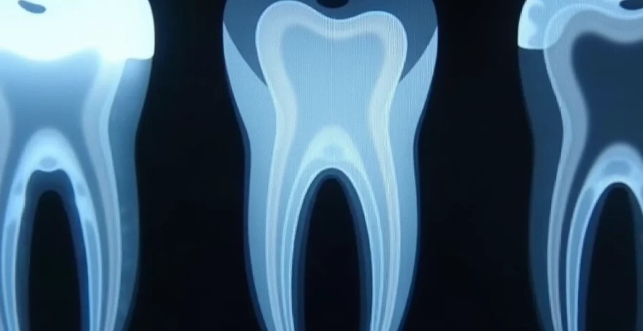
Pulp chamber morphology and root canal visibility
The pulp chamber and root canal system appear as radiolucent spaces within the tooth structure, representing the soft tissue components containing nerves, blood vessels, and connective tissue. In healthy teeth, these spaces should demonstrate age-appropriate dimensions, with younger patients typically showing larger pulp chambers that gradually reduce in size due to secondary dentine deposition over time.
Root canal morphology varies significantly between different tooth types, with incisors typically displaying single, well-defined canals, whilst molars may present complex multi-canal systems. The pulp chamber outline should appear smooth and continuous, without any irregular enlargements or constrictions that might indicate pathological processes such as internal resorption or calcific metamorphosis.
Periodontal ligament space width and lamina dura integrity
The periodontal ligament space appears as a thin, radiolucent line surrounding the entire root surface, maintaining relatively uniform width throughout its length. This space typically measures between 0.15-0.25 millimetres in healthy individuals, though slight variations may occur depending on functional demands and individual anatomical factors. Any significant widening or irregularity of this space may indicate periodontal pathology or orthodontic movement.
Adjacent to the periodontal ligament space, the lamina dura presents as a thin, radiopaque line representing the alveolar bone proper. This structure should appear continuous and well-defined around the entire root perimeter, maintaining consistent density and thickness. Disruptions or loss of lamina dura continuity often signify pathological processes affecting the periapical tissues.
Alveolar bone trabecular pattern recognition
Healthy alveolar bone demonstrates a characteristic trabecular pattern consisting of fine, interconnected bony trabeculae separated by marrow spaces. The trabecular architecture varies between individuals and anatomical locations, with the mandible typically showing coarser patterns compared to the maxilla. This variation reflects differences in functional demands and bone density between various oral regions.
The trabecular pattern should appear uniform throughout the alveolar process, without areas of increased radiolucency that might suggest bone loss or cystic changes. Normal bone density variations occur with age, with older patients typically demonstrating more pronounced trabecular patterns due to increased mineralisation and reduced marrow space volume.
Cortical bone boundaries and mandibular canal visualisation
Cortical bone boundaries appear as continuous, radiopaque lines that define the outer margins of the alveolar process and jaw bones. These structures should maintain consistent thickness and density, providing clear demarcation between bone and adjacent soft tissues. The cortical plates serve as important landmarks for assessing bone health and planning surgical procedures.
The mandibular canal presents as a well-defined, radiolucent structure coursing through the mandibular body, housing the inferior alveolar neurovascular bundle. This anatomical landmark should appear as a continuous, smooth-walled channel with clearly visible cortical borders. Proper identification of the mandibular canal is crucial for surgical planning and avoiding complications during invasive procedures.
Optimal radiographic technique parameters for diagnostic quality
Achieving consistently high-quality dental radiographs requires meticulous attention to technical parameters and positioning protocols. The diagnostic value of any radiographic examination depends heavily on proper technique execution, as even subtle variations in positioning or exposure settings can significantly impact image quality and diagnostic accuracy. Understanding the principles underlying optimal radiographic technique enables practitioners to produce images that faithfully represent the anatomical structures under examination.
Bitewing x-ray positioning for interproximal caries detection
Bitewing radiographs serve as the gold standard for detecting interproximal caries and assessing crestal bone levels. Proper positioning requires precise alignment of the X-ray beam perpendicular to the film or sensor, with the bite tab positioned to capture both maxillary and mandibular posterior teeth simultaneously. The horizontal angulation must be adjusted to ensure contact areas between adjacent teeth appear open, preventing overlap that could obscure early carious lesions.
The vertical angulation for bitewing projections typically ranges between +5 to +15 degrees, depending on the patient’s occlusal plane orientation. This slight positive angulation helps compensate for the natural curve of Spee and ensures optimal visualisation of the cervical regions where caries frequently develop. Consistent positioning protocols are essential for producing comparable images during serial examinations, facilitating accurate monitoring of disease progression or treatment outcomes.
Periapical film placement using paralleling technique
The paralleling technique represents the preferred method for acquiring periapical radiographs due to its ability to produce images with minimal geometric distortion. This approach utilises film or sensor holders that maintain the receptor parallel to the long axis of the tooth whilst positioning the X-ray source at an adequate distance to ensure parallel beam geometry. The technique requires careful attention to receptor placement, ensuring adequate coverage of the apex whilst avoiding patient discomfort.
Proper paralleling technique execution involves positioning the receptor approximately 6-8 millimetres lingual to the teeth of interest, with the X-ray beam directed perpendicular to both the tooth and the receptor. This geometric arrangement produces images with accurate dimensional relationships and minimal magnification, facilitating precise measurements for endodontic and surgical procedures. The use of beam-indicating devices further enhances positioning accuracy and reduces radiation exposure to surrounding tissues.
Panoramic radiograph patient positioning protocols
Panoramic radiography requires precise patient positioning to achieve optimal image quality across the entire maxillofacial region. The patient’s head must be positioned with the Frankfurt horizontal plane parallel to the floor, whilst the midsagittal plane remains perpendicular to the film plane. Proper chin positioning is crucial, as excessive chin elevation or depression can result in significant image distortion and loss of diagnostic information.
The patient’s bite should be positioned on the bite guide with the teeth slightly apart, typically maintaining a 2-3 millimetre separation. This positioning prevents superimposition of the cervical spine over the mandibular anterior region whilst ensuring optimal visualisation of the temporomandibular joint areas. Additionally, the tongue should be positioned against the palate to eliminate the air space that would otherwise appear as a radiolucent band across the maxillary region.
Digital sensor exposure settings and kvp optimisation
Digital radiographic systems require careful calibration of exposure parameters to achieve optimal image quality whilst minimising radiation dose. The kilovoltage peak (kVp) setting directly influences image contrast and penetration capability, with most dental applications utilising ranges between 60-70 kVp for intraoral projections. Higher kVp values provide greater penetration and reduced contrast, whilst lower values enhance contrast at the expense of increased patient dose.
Sensor sensitivity characteristics vary between manufacturers and should be considered when establishing exposure protocols. Most modern digital sensors demonstrate significantly higher sensitivity compared to conventional film, often requiring exposure reductions of 50-80% whilst maintaining diagnostic quality. Regular calibration and quality assurance procedures ensure consistent image quality and optimal dose utilisation throughout the sensor’s operational lifespan.
Pathology-free radiographic indicators
Recognising the absence of pathological changes represents a fundamental skill in radiographic interpretation, requiring thorough knowledge of normal anatomical variations and their typical presentation. Healthy dental structures exhibit predictable radiographic characteristics that serve as baseline references for identifying disease processes. The systematic evaluation of specific anatomical regions ensures comprehensive assessment and reduces the likelihood of overlooking subtle pathological changes that may require intervention.
Absence of radiolucent lesions in apical regions
Healthy periapical tissues demonstrate uniform bone density without radiolucent areas that might suggest inflammatory or infectious processes. The bone immediately surrounding root apices should exhibit normal trabecular patterns consistent with adjacent areas, maintaining continuity of the lamina dura around the entire root perimeter. The absence of periapical radiolucencies indicates vital pulp tissues and healthy periapical supporting structures.
When evaluating periapical regions, particular attention should be paid to the root apex morphology and surrounding bone architecture. Normal root apices appear well-formed with complete root development, whilst the surrounding bone demonstrates mature trabecular patterns without areas of sclerosis or rarefaction. Any deviation from these normal patterns may indicate previous trauma, infection, or developmental anomalies requiring further investigation.
Intact interproximal contact points without carious lesions
Healthy interproximal surfaces appear as continuous, uninterrupted radiopaque areas where adjacent teeth contact. The enamel surfaces should maintain their characteristic high-density appearance without any radiolucent areas that might indicate demineralisation or carious involvement. Contact areas should appear tight and well-defined, preventing food impaction and bacterial accumulation that could predispose to carious development.
The cervical regions of interproximal surfaces require careful evaluation, as these areas represent common sites for caries initiation due to plaque accumulation and reduced self-cleansing action. In healthy dentition, the cervical enamel maintains uniform density extending to the cemento-enamel junction, where it transitions smoothly to the root surface without any notching or irregularities that might suggest carious involvement or cervical resorption.
Uniform bone density patterns in maxillary and mandibular arches
Healthy alveolar bone exhibits consistent density patterns throughout both dental arches, with gradual variations that reflect normal anatomical differences between various oral regions. The maxillary bone typically demonstrates finer trabecular patterns compared to the mandible, reflecting differences in bone density and functional demands. Uniform bone density indicates adequate mineralisation and normal metabolic activity within the alveolar process.
The crestal bone levels should maintain appropriate heights relative to the cemento-enamel junctions of adjacent teeth, typically positioned within 2-3 millimetres of these anatomical landmarks. Normal crestal bone appears as a sharp, radiopaque line connecting adjacent lamina dura, maintaining consistent height and density throughout the dental arch. Any significant variations in crestal bone levels may indicate periodontal disease or localised bone loss requiring therapeutic intervention.
Normal Crown-to-Root ratio assessment
Healthy teeth demonstrate appropriate crown-to-root proportions that reflect normal development and adequate periodontal support. Most teeth exhibit crown-to-root ratios ranging from 1:1.5 to 1:2, depending on the specific tooth type and individual anatomical variations. These proportions provide essential information regarding the tooth’s ability to withstand functional loads and long-term prognosis under various treatment scenarios.
Root morphology should appear well-developed with appropriate length and thickness to support the overlying crown structure. Single-rooted teeth typically demonstrate conical root forms with gradual tapering towards the apex, whilst multi-rooted teeth show well-separated roots with distinct anatomical characteristics. Proper root development indicates normal tooth formation and adequate foundation for long-term function and stability.
Age-related radiographic variations in healthy dentition
Understanding normal age-related changes in dental radiographs is essential for accurate interpretation and avoiding misdiagnosis of physiological processes as pathological conditions. As individuals age, various structural modifications occur within dental tissues, alveolar bone, and supporting structures that reflect normal adaptation to functional demands and cellular activity. These changes follow predictable patterns that can be distinguished from disease processes through careful radiographic analysis and clinical correlation.
Pulp chamber dimensions undergo progressive reduction throughout life due to continuous secondary dentine deposition, a process known as pulpal sclerosis. Young patients typically display large pulp chambers with prominent pulp horns, whilst older individuals show significantly reduced pulpal spaces with calcified areas that may appear as radiopaque deposits within the chamber. This physiological process should not be confused with pathological calcifications resulting from trauma or pulpal inflammation.
The trabecular bone pattern becomes increasingly coarse and well-defined with advancing age, reflecting changes in bone remodelling activity and mineral density. Older patients often demonstrate more pronounced trabecular architecture with larger marrow spaces, creating a characteristic “cotton wool” appearance in some regions. These age-related bone changes represent normal adaptations to altered functional demands and should be distinguished from pathological bone loss or metabolic disorders affecting bone quality.
Root resorption may occur as a normal physiological process in primary dentition during the transition to permanent teeth, but minimal root shortening can also be observed in permanent teeth of elderly patients. This physiological root resorption typically affects the apical third of roots and progresses slowly over extended periods. The process should be differentiated from pathological external resorption resulting from trauma, orthodontic movement, or inflammatory conditions that typically demonstrate more rapid progression and irregular resorption patterns.
Common radiographic artefacts versus pathological findings
Distinguishing between radiographic artefacts and genuine pathological findings represents one of the most challenging aspects of dental radiographic interpretation. Artefacts can arise from various sources including improper technique, processing errors, or patient-related factors, and may closely mimic pathological conditions leading to potential misdiagnosis. Developing the ability to recognise and differentiate these imaging anomalies from actual disease processes is crucial for accurate diagnosis and appropriate treatment planning.
Processing artefacts in digital radiography can create artificial radiolucencies or radiopacities that may simulate carious lesions or restorative materials. Sensor contamination or damage can produce consistent patterns across multiple images, whilst software processing errors may create geometric distortions or density variations that appear pathological. Regular quality assurance procedures and equipment maintenance help minimise these technical artefacts and ensure consistent image quality.
Patient movement during exposure creates characteristic blur patterns that can obscure anatomical details and potentially mask pathological changes. Unlike pathological findings that maintain consistent relationships to anatomical structures, movement artefacts typically affect the entire image uniformly and can be recognised by their characteristic appearance. Proper patient stabilisation and clear instructions regarding the need to remain motionless during exposure help prevent these image degradation issues.
Overlapping anatomical structures can create complex radiographic patterns that may be mistaken for pathological changes, particularly in panoramic radiographs where multiple anatomical planes are represented simultaneously. The cervical spine, hyoid bone, and soft tissue shadows can superimpose over dental structures, creating apparent radiolucencies or radiopacities that have no pathological significance. Understanding normal anatomical relationships and their radiographic appearance helps differentiate these normal structures from pathological findings requiring intervention.