The accumulation of excess fat around the midsection represents far more than a cosmetic concern—it signals a cascade of potentially life-threatening metabolic disruptions occurring deep within your body. While society often focuses on the visible aspects of weight gain, the most dangerous fat deposits lie hidden beneath the surface, silently wreaking havoc on vital organ systems. This visceral adipose tissue, nestled around your liver, pancreas, and intestines, operates as a metabolically active endocrine organ, secreting inflammatory compounds and disrupting hormonal balance in ways that can fundamentally alter your health trajectory.
Modern research has unveiled the stark reality that abdominal obesity serves as a primary driver of chronic disease development, with implications extending far beyond traditional cardiovascular concerns. The intricate web of physiological changes triggered by excess visceral fat creates a perfect storm of metabolic dysfunction, inflammatory processes, and hormonal imbalances that can accelerate aging and disease progression at the cellular level.
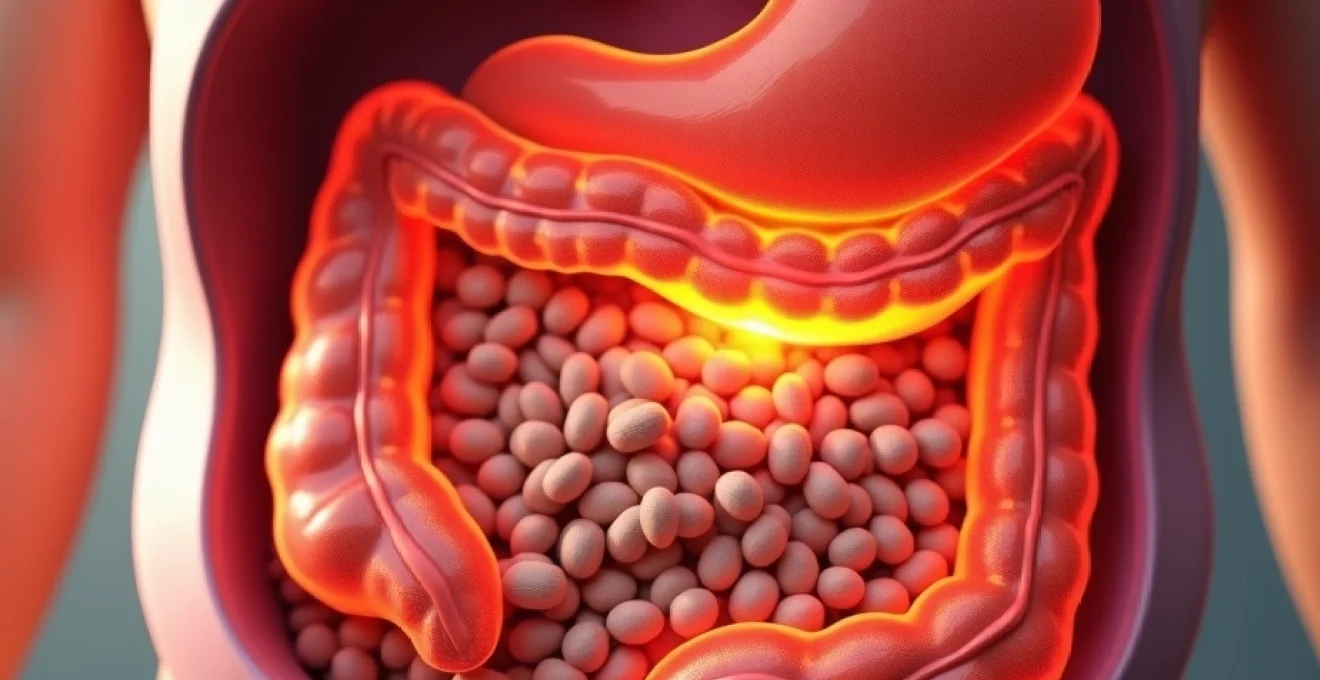
Visceral adipose tissue: understanding central obesity’s pathophysiology
Visceral adipose tissue represents a distinct anatomical and functional entity that differs dramatically from the subcutaneous fat most people can observe and measure. This metabolically active tissue forms a complex network around your abdominal organs, creating what researchers describe as an inflammatory microenvironment that can influence systemic health in profound ways.
Intra-abdominal fat distribution patterns and metabolic consequences
The distribution of intra-abdominal fat follows specific anatomical patterns that directly correlate with metabolic risk profiles. Omental fat, which drapes over the intestines like an apron, tends to accumulate more readily in individuals with insulin resistance and represents the most metabolically dangerous depot. This particular fat distribution pattern creates a direct pathway for inflammatory compounds to enter portal circulation, affecting liver function and glucose metabolism immediately.
Mesenteric fat, located between the layers of tissue that support the intestines, contributes significantly to the overall visceral fat burden and plays a crucial role in inflammatory cascade activation . The proximity of these fat deposits to major blood vessels means that the toxic substances they produce can rapidly enter systemic circulation, creating widespread metabolic disruption throughout the body.
Visceral fat versus subcutaneous fat: cellular composition differences
The cellular architecture of visceral adipose tissue differs markedly from subcutaneous deposits in ways that explain its heightened metabolic activity. Visceral fat contains a higher concentration of large adipocytes, which are more prone to inflammatory responses and insulin resistance development. These enlarged fat cells become dysfunctional, losing their ability to properly store and release fatty acids in response to metabolic demands.
Furthermore, visceral fat demonstrates increased vascularization and innervation compared to subcutaneous deposits, facilitating rapid communication with the central nervous system and enhanced responsiveness to stress hormones like cortisol. This heightened sensitivity creates a feedback loop where stress contributes to visceral fat accumulation, which in turn amplifies stress-related metabolic disruptions.
Waist-to-hip ratio classification systems and clinical thresholds
Healthcare professionals utilize specific measurement protocols to assess visceral fat accumulation and associated health risks. The waist circumference measurement, taken at the midpoint between the lowest rib and the iliac crest, provides the most practical clinical assessment tool. For women, measurements exceeding 80cm indicate increased health risks, while men face elevated risks with waist measurements above 94cm.
The waist-to-hip ratio offers additional insights into fat distribution patterns, with ratios above 0.85 for women and 0.90 for men suggesting android or “apple-shaped” obesity patterns associated with higher cardiovascular and metabolic risks. These measurements correlate strongly with visceral fat volume as determined by more sophisticated imaging techniques.
Computed tomography and MRI imaging for visceral fat quantification
Advanced imaging techniques provide precise quantification of visceral adipose tissue volume, offering researchers and clinicians detailed insights into fat distribution patterns. CT scans at the L4-L5 vertebral level can accurately distinguish between visceral and subcutaneous fat compartments, with visceral fat areas exceeding 130 cm² in men and 110 cm² in women indicating significantly elevated health risks.
MRI imaging offers superior soft tissue contrast without radiation exposure, enabling detailed analysis of fat distribution throughout the abdominal cavity. These techniques have revealed that individuals with identical BMI measurements can have dramatically different visceral fat volumes, explaining why some people with “normal” weights still face significant metabolic health risks.
Cardiovascular disease risk amplification through abdominal adiposity
The relationship between abdominal fat accumulation and cardiovascular disease represents one of the most well-established connections in modern medicine. Visceral adipose tissue creates a perfect storm of conditions that accelerate atherosclerosis development and increase the likelihood of cardiac events through multiple interconnected pathways. The inflammatory compounds produced by visceral fat directly damage blood vessel walls, while simultaneously promoting blood clot formation and disrupting normal vascular function.
Coronary artery disease progression in central obesity patients
Individuals with central obesity experience accelerated coronary artery disease progression through several distinct mechanisms. The chronic inflammatory state created by visceral fat promotes endothelial dysfunction, reducing the ability of coronary arteries to dilate properly in response to increased oxygen demands. This impairment can lead to myocardial ischemia even during routine daily activities.
Additionally, the altered lipid profile commonly associated with abdominal obesity—characterized by elevated triglycerides, reduced HDL cholesterol, and increased small, dense LDL particles—creates an atherogenic environment that accelerates plaque formation within coronary vessels. These metabolic changes can begin years before clinical symptoms become apparent, making early intervention crucial.
Hypertension development via Renin-Angiotensin-Aldosterone system activation
Visceral adipose tissue directly influences blood pressure regulation through its effects on the renin-angiotensin-aldosterone system (RAAS). Fat cells within the abdominal cavity produce angiotensinogen, a precursor to angiotensin II, which promotes vasoconstriction and sodium retention. This local production of RAAS components creates a microenvironment that favors hypertension development.
The mechanical compression effects of excess abdominal fat also contribute to elevated blood pressure by reducing kidney function efficiency. As visceral fat accumulates around the kidneys, it can impair their ability to regulate fluid balance and blood pressure effectively, creating a cycle where obesity promotes hypertension, which in turn accelerates cardiovascular damage.
Atherosclerotic plaque formation and inflammatory cytokine release
The inflammatory cytokines released by visceral fat—including tumor necrosis factor-alpha, interleukin-6, and C-reactive protein—play direct roles in atherosclerotic plaque formation and destabilization. These compounds promote the migration of immune cells into arterial walls, where they contribute to foam cell formation and plaque development. The result is an accelerated atherosclerotic process that can affect multiple vascular beds simultaneously.
Perhaps more concerning is the role of these inflammatory mediators in plaque destabilization. The cytokines produced by visceral fat can weaken the fibrous caps that stabilize atherosclerotic plaques, increasing the likelihood of plaque rupture and subsequent thrombotic events such as heart attacks and strokes.
Cardiac output strain and left ventricular hypertrophy mechanisms
The increased metabolic demands of excess visceral adipose tissue place significant strain on cardiac function. The heart must work harder to perfuse the enlarged fat mass, leading to increased cardiac output requirements and elevated filling pressures. Over time, this chronic workload increase can result in left ventricular hypertrophy, a condition that impairs the heart’s ability to relax properly between beats.
The development of diastolic dysfunction often precedes systolic heart failure in individuals with abdominal obesity. This progression represents a continuum where initially compensatory cardiac adaptations eventually become maladaptive, leading to reduced exercise tolerance, fluid retention, and ultimately, heart failure symptoms.
Type 2 diabetes mellitus onset through insulin resistance pathways
The connection between abdominal fat accumulation and type 2 diabetes development represents one of the most direct and devastating consequences of visceral obesity. Unlike subcutaneous fat, which can expand relatively benignly to accommodate excess energy storage, visceral adipose tissue quickly becomes dysfunctional when overwhelmed. This dysfunction triggers a cascade of metabolic disruptions that systematically undermine your body’s ability to maintain healthy blood glucose levels.
The process begins at the cellular level, where enlarged visceral fat cells lose their ability to respond appropriately to insulin signals. These dysfunctional adipocytes begin releasing free fatty acids inappropriately, flooding the bloodstream with lipids that interfere with glucose uptake in muscle and liver tissues. Simultaneously, these same fat cells reduce their production of beneficial compounds like adiponectin , which normally helps maintain insulin sensitivity throughout the body.
The liver becomes a primary target of this metabolic disruption, as it receives a concentrated dose of inflammatory compounds and excess fatty acids directly from visceral fat through the portal circulation. This toxic exposure promotes hepatic insulin resistance and triggers increased glucose production, even when blood sugar levels are already elevated. The result is a relentless cycle where the liver produces more glucose while peripheral tissues become increasingly unable to utilize it effectively.
Research indicates that individuals with excess visceral fat are up to five times more likely to develop type 2 diabetes compared to those with predominantly subcutaneous fat distribution, regardless of overall body weight.
The timeline of diabetes development in visceral obesity follows a predictable pattern that can span years or even decades. Initially, the pancreas compensates for increasing insulin resistance by producing more insulin, maintaining normal blood glucose levels at the cost of chronically elevated insulin concentrations. This hyperinsulinemic state contributes to further fat accumulation, particularly in the abdominal region, creating a self-perpetuating cycle of metabolic dysfunction.
Inflammatory cascade activation and cytokine storm phenomena
Visceral adipose tissue functions as an inflammatory organ, producing a constant stream of pro-inflammatory cytokines that create a state of chronic low-grade inflammation throughout the body. This inflammatory environment affects virtually every organ system, accelerating aging processes and increasing susceptibility to numerous chronic diseases. The key inflammatory mediators include tumor necrosis factor-alpha (TNF-α), interleukin-6 (IL-6), and C-reactive protein (CRP), each contributing unique pathogenic effects.
The inflammatory cascade begins when visceral fat cells become stressed due to their enlarged size and altered metabolic environment. These stressed adipocytes attract immune cells called macrophages, which infiltrate the fat tissue and establish a chronic inflammatory state. Unlike the acute inflammation that helps fight infections, this chronic inflammatory condition serves no protective purpose and instead damages healthy tissues throughout the body.
TNF-α represents one of the most potent inflammatory cytokines produced by visceral fat, directly interfering with insulin signaling pathways and promoting further fat accumulation. This compound can cross the blood-brain barrier, affecting hypothalamic function and disrupting normal appetite regulation. The result is often increased food cravings and reduced satiety signals, making weight management increasingly difficult.
Interleukin-6 contributes to the inflammatory environment while simultaneously promoting the production of acute-phase proteins in the liver. Elevated IL-6 levels correlate strongly with increased cardiovascular disease risk and have been implicated in the development of depression and cognitive decline. The systemic effects of this cytokine demonstrate how abdominal obesity can influence mental health and neurological function.
C-reactive protein serves as a marker of systemic inflammation and independently predicts cardiovascular events. Individuals with excess visceral fat often maintain CRP levels that are two to three times higher than those with healthy fat distribution patterns. This elevation indicates a chronic inflammatory state that accelerates atherosclerosis and increases the risk of blood clot formation.
Hormonal disruption mechanisms in abdominal fat accumulation
The hormonal disruptions associated with visceral fat accumulation create a complex web of metabolic dysfunction that extends far beyond simple energy balance equations. Abdominal obesity fundamentally alters the production, distribution, and effectiveness of numerous hormones that regulate metabolism, stress response, appetite, and energy expenditure. Understanding these hormonal changes is crucial for comprehending why visceral fat accumulation becomes increasingly difficult to reverse once established.
Cortisol dysregulation and Hypothalamic-Pituitary-Adrenal axis dysfunction
Chronic stress and elevated cortisol levels play pivotal roles in promoting visceral fat accumulation through multiple mechanisms. The hypothalamic-pituitary-adrenal (HPA) axis becomes dysregulated in individuals with abdominal obesity, leading to altered cortisol production patterns and increased tissue sensitivity to stress hormones. This dysregulation creates a vicious cycle where stress promotes visceral fat accumulation, which in turn amplifies stress hormone production.
Cortisol directly stimulates the differentiation of pre-adipocytes into mature fat cells, particularly in the abdominal region where adipose tissue contains higher concentrations of cortisol receptors. Additionally, elevated cortisol levels promote increased appetite and cravings for high-calorie, high-fat foods through their effects on brain reward pathways. The enzyme 11β-hydroxysteroid dehydrogenase type 1, which converts inactive cortisone to active cortisol, is particularly abundant in visceral fat, creating a local environment of heightened stress hormone activity.
Leptin resistance development and satiety signal impairment
Leptin, often called the “satiety hormone,” normally signals the brain when energy stores are adequate, helping to regulate food intake and energy expenditure. However, individuals with excess visceral fat frequently develop leptin resistance, where the brain becomes insensitive to leptin’s appetite-suppressing signals despite elevated circulating levels of the hormone. This resistance contributes to persistent hunger and reduced metabolic rate, making weight loss increasingly challenging.
The inflammatory cytokines produced by visceral fat interfere with leptin signaling pathways in the hypothalamus, disrupting the normal feedback mechanisms that maintain energy balance. Additionally, elevated triglyceride levels associated with abdominal obesity can impair leptin transport across the blood-brain barrier, further reducing the effectiveness of this crucial metabolic hormone. The result is a state where the body continues to promote food intake and fat storage despite already excessive energy reserves.
Adiponectin reduction and anti-inflammatory protection loss
Adiponectin represents one of the few beneficial compounds produced by fat tissue, offering anti-inflammatory and insulin-sensitizing effects that help maintain metabolic health. However, visceral fat produces significantly less adiponectin compared to subcutaneous fat, and adiponectin levels tend to decline as abdominal obesity develops. This reduction eliminates an important protective factor against metabolic dysfunction and cardiovascular disease.
The loss of adiponectin’s protective effects contributes to increased insulin resistance, enhanced inflammatory responses, and impaired fat oxidation in muscle tissue. Low adiponectin levels also correlate with increased risk of developing metabolic syndrome, type 2 diabetes, and cardiovascular disease. The decline in this beneficial hormone represents another mechanism by which visceral fat accumulation perpetuates metabolic dysfunction and disease risk.
Growth hormone deficiency and lipolysis inhibition
Growth hormone plays crucial roles in maintaining healthy body composition by promoting muscle development and facilitating fat breakdown through lipolysis. Individuals with excess visceral fat often experience reduced growth hormone production and altered growth hormone release patterns, particularly during sleep. This deficiency contributes to further muscle loss and impaired fat metabolism, creating conditions that favor continued abdominal fat accumulation.
The inflammatory environment created by visceral fat interferes with growth hormone’s metabolic effects, reducing its ability to promote fat oxidation and muscle protein synthesis. Additionally, insulin resistance associated with abdominal obesity can suppress growth hormone-releasing hormone production, further compromising this important metabolic hormone. The result is a hormonal environment that favors fat storage over fat burning and promotes the loss of metabolically active muscle tissue.
Non-alcoholic fatty liver disease progression and hepatic steatosis
The liver bears the brunt of visceral fat’s toxic effects due to its unique anatomical position downstream from abdominal fat deposits through the portal circulation system. This direct exposure to inflammatory compounds and excess fatty acids makes the liver particularly vulnerable to developing non-alcoholic fatty liver disease (NAFLD), a condition that affects up to 80% of individuals with significant abdominal obesity. The progression from simple hepatic steatosis to more severe forms of liver disease represents one of the most concerning consequences of visceral fat accumulation.
The development of NAFLD begins when visceral fat releases excessive
amounts of free fatty acids directly into the portal circulation, overwhelming the liver’s capacity to process these lipids effectively. Under normal circumstances, the liver can handle moderate amounts of fatty acids by converting them into triglycerides for storage or oxidizing them for energy. However, the continuous influx from dysfunctional visceral fat creates a state of lipotoxicity where the liver becomes saturated with fat, leading to hepatocyte dysfunction and inflammatory activation.
The progression from simple steatosis to non-alcoholic steatohepatitis (NASH) occurs when accumulated liver fat triggers inflammatory responses and oxidative stress. This transition marks a critical point where reversible fat accumulation becomes potentially irreversible liver damage, including fibrosis and cirrhosis. The inflammatory cytokines produced by visceral fat, particularly TNF-α and IL-6, directly contribute to hepatic inflammation and stellate cell activation, promoting collagen deposition and scarring within liver tissue.
Hepatic insulin resistance develops as a direct consequence of this fatty infiltration and inflammatory activation. The liver loses its ability to respond appropriately to insulin signals, leading to continued glucose production even when blood sugar levels are elevated. This hepatic glucose overproduction contributes significantly to the development of type 2 diabetes and makes blood sugar control increasingly difficult in affected individuals.
The diagnostic criteria for NAFLD include hepatic steatosis affecting more than 5% of hepatocytes, as determined by imaging studies or liver biopsy, in the absence of significant alcohol consumption. Advanced imaging techniques such as magnetic resonance spectroscopy can detect hepatic fat content as low as 1%, enabling earlier detection and intervention. The condition affects an estimated 25% of the global population, with prevalence rates reaching 80-90% among individuals with obesity and type 2 diabetes.
Studies demonstrate that individuals with visceral obesity have a 75% likelihood of developing some degree of hepatic steatosis, with 20-30% progressing to more advanced forms of liver disease within a decade.
The metabolic consequences of NAFLD extend beyond liver function, creating systemic effects that accelerate cardiovascular disease development and worsen insulin resistance throughout the body. The liver’s impaired ability to synthesize beneficial proteins like albumin and transport lipoproteins disrupts normal lipid metabolism, leading to atherogenic dyslipidemia characterized by elevated triglycerides and reduced HDL cholesterol levels. These changes create an environment that promotes atherosclerosis and increases the risk of cardiovascular events.
Treatment strategies for NAFLD focus primarily on addressing the underlying visceral obesity and metabolic dysfunction that drive the condition. Weight loss of 5-10% can significantly reduce hepatic steatosis, while losses exceeding 10% may reverse inflammatory changes and early fibrosis. The Mediterranean diet pattern, characterized by high consumption of olive oil, fish, vegetables, and whole grains, has demonstrated particular efficacy in reducing liver fat content and improving hepatic insulin sensitivity.
The prognosis for individuals with NAFLD varies significantly based on disease stage at diagnosis and the effectiveness of lifestyle interventions. Simple steatosis carries a relatively benign prognosis with appropriate management, while NASH with fibrosis represents a more serious condition that can progress to cirrhosis and hepatocellular carcinoma. Regular monitoring through imaging studies and liver function tests enables healthcare providers to track disease progression and adjust treatment strategies accordingly.